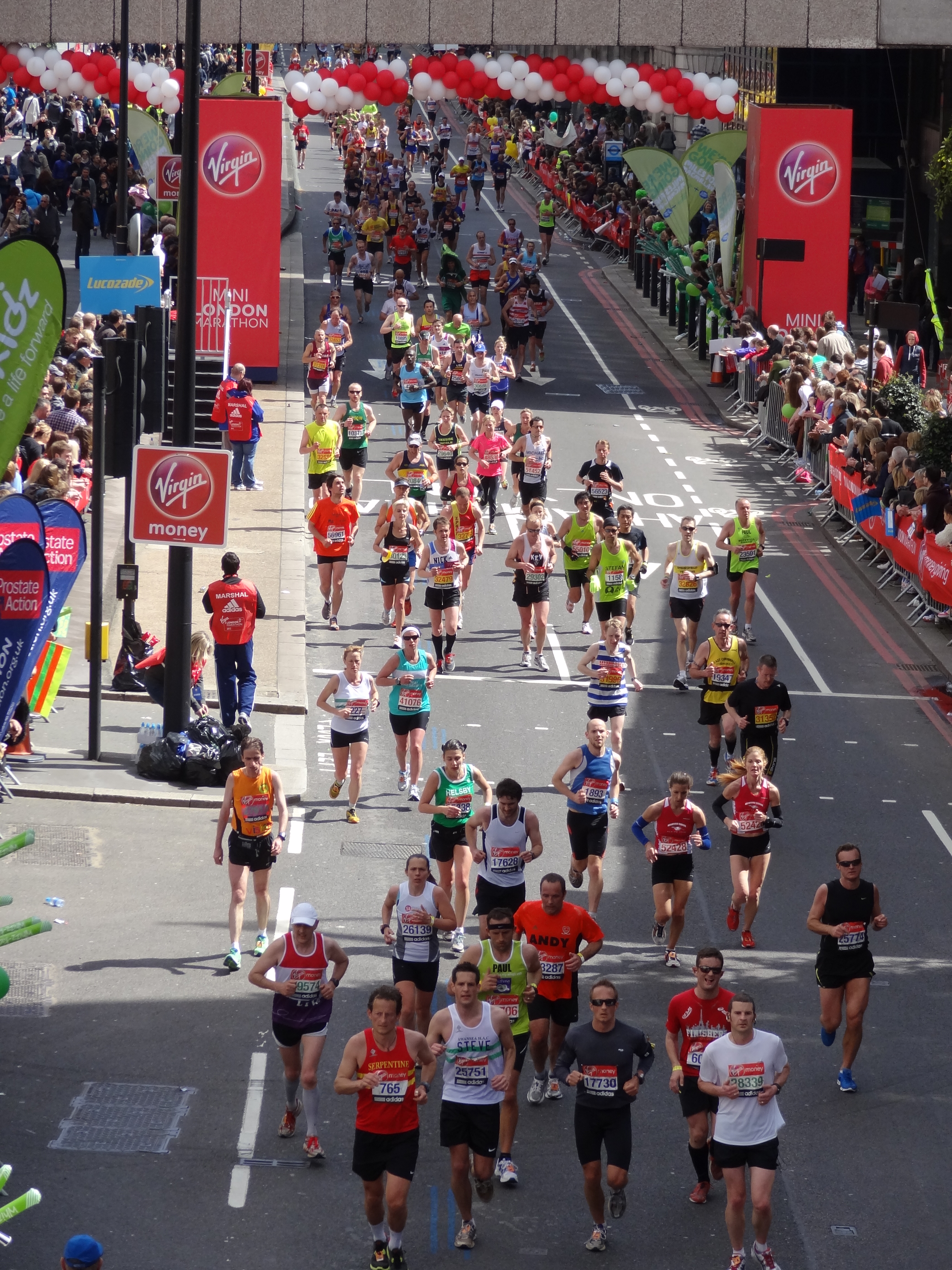
Given the significance of this year's Virgin London Marathon, with several athletes attempting to achieve a qualification time for the 2012 London Olympics & the fact that I have had the pleasure to work with a handful of those runners in recent days, weeks & months, I decided to spend Sunday in London to add my support to the thousands of others lining the streets. Surprisingly, I have never stood along the course to watch before, despite being involved in other capacities on one or two previous occasions & I was really impressed with the atmosphere. Away from the elite athletes, the thousands of people running for charities, personal causes & passions is incredible & it is incredibly moving to see the support offered to those driven individuals.
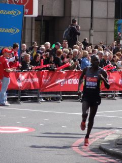
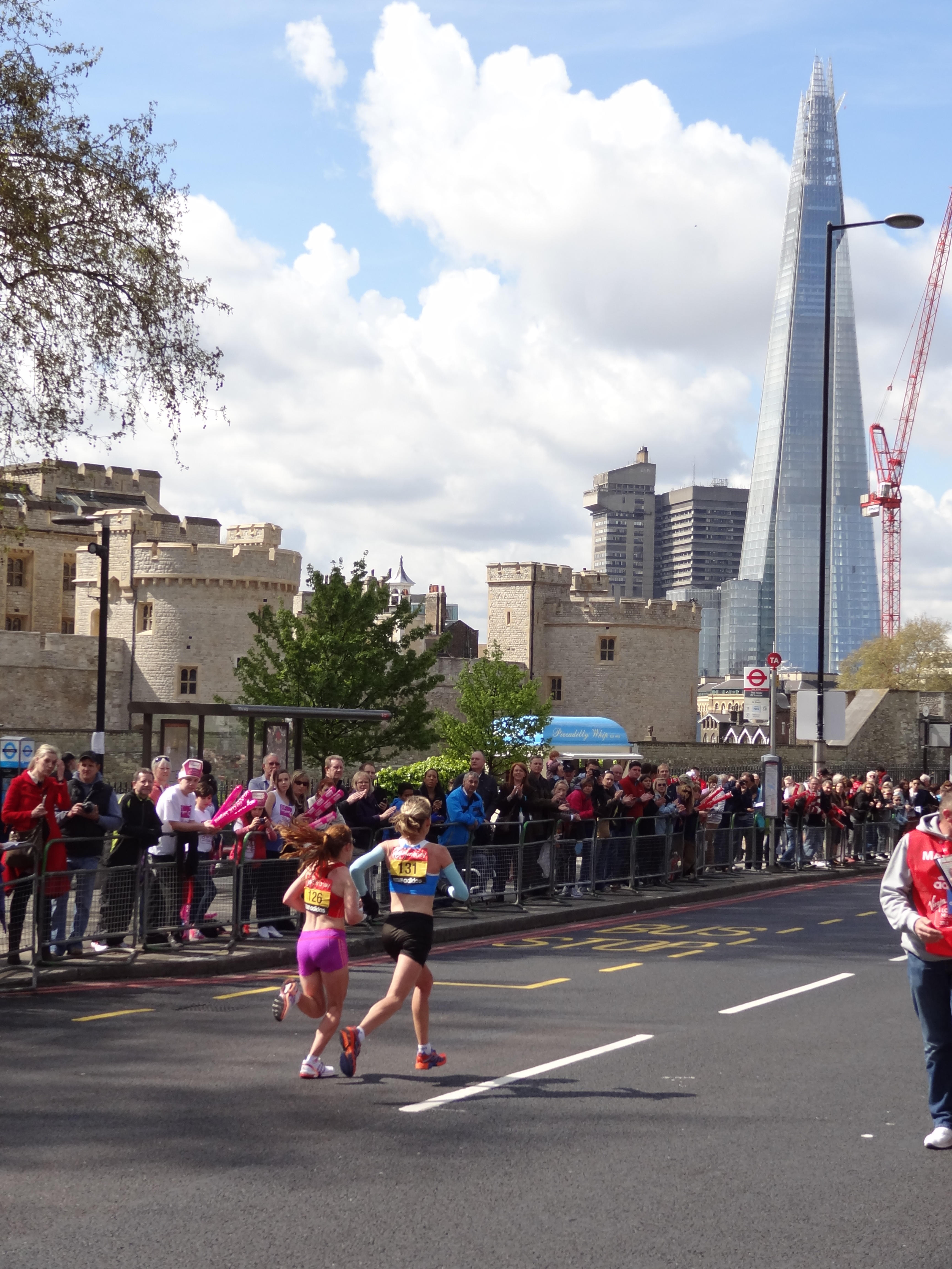
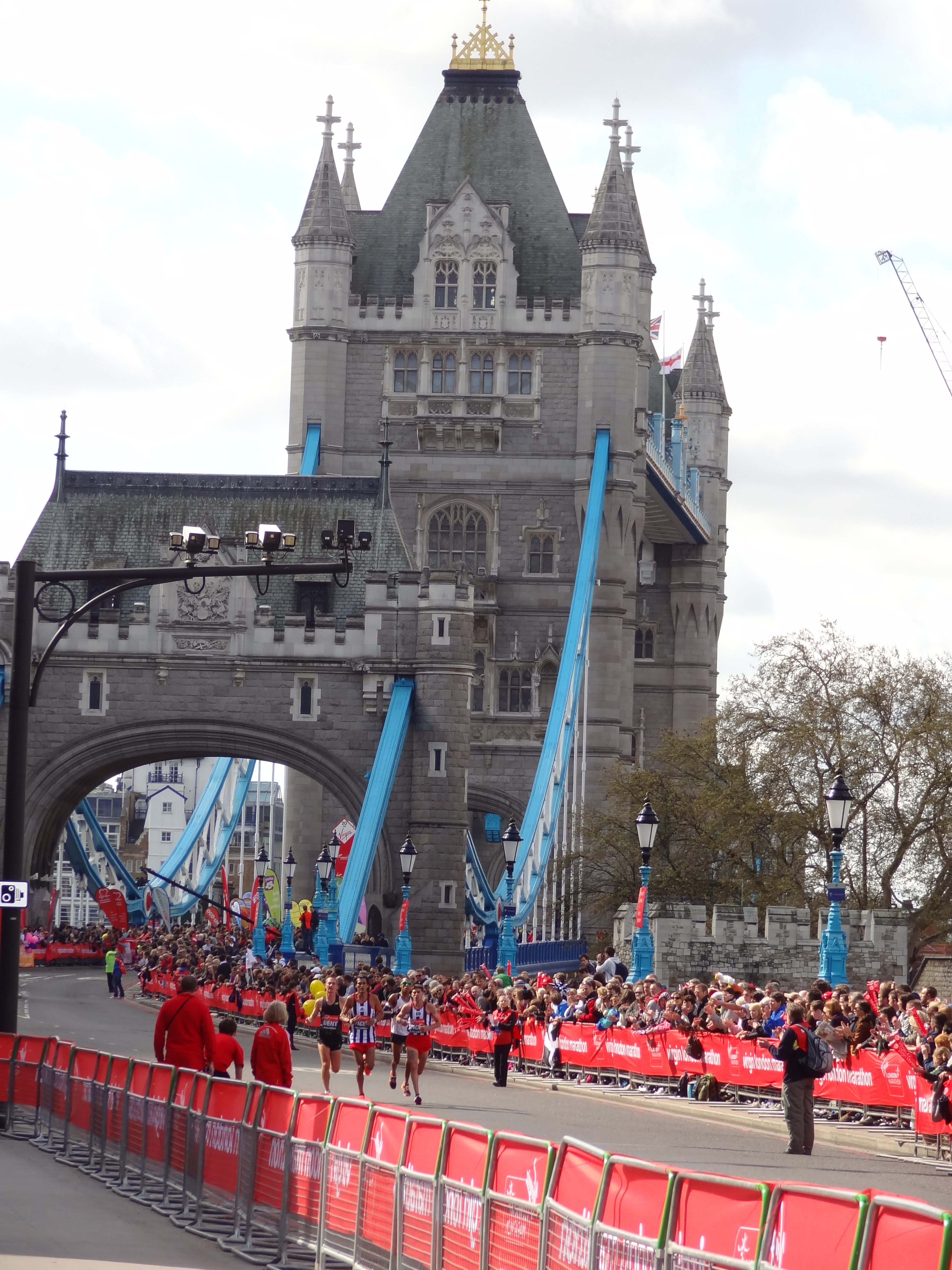
I picked myself a couple of vantage points, initially skirting with the pernickety health & safety laws governing this country at present, to perch momentarily (I was asked to refrain from climbing) on the side of Tower Bridge at around mile 15, then moving to mile 22 to watch in greater comfort. I was fortunate enough to have picked my spot just before the wheelchair athletes came into view, followed shortly by the elite women & then the elite men.
After the elite, came the club runners heading up the field of around 36,000 participants, some of whom will take more than just one day to finish the race. One such entrant, Clare Lomas, will take around 3 weeks to complete the course, wearing a "Rewalk Suit", which is helping her mobilise despite being paralysed after sustaining spinal injuries in a horse riding accident. Other notable achievements were that of Fauja Singh, the oldest marathon runner at 101, who completed the event in seven hours & 49 minutes; Edward Lumley who, in clocking two hours 59 minutes, broke the world record for the fastest marathon dressed as a vegetable & Sasha Kenney, who raised £2,000 for the NSPCC by hula hooping the course in five hours & 5 minutes.
The elite races were won by Kenyans Wilson Kipsang & Mary Keitany, stopping the clock in 2:04.44 & 2:18.37 respectively. Neither time was fast enough for a course record, but one athlete who did manage to achieve a record was David Weir, who won the men's wheelchair race for a record-equalling 6th time, an achievement previously attained by Baroness Tanni Grey-Thompson.
Whilst David battled out a sprint finish to cross the line just a second ahead of Switzerland's Hug, Shelley Woods dominated the women's wheelchair race with over 3 minutes separating her from Tsuchida (Japan) in second place.
Unfortunately, Team GB's dominance in the wheelchair events was not to be matched by the able bodied Brits. Top-finishing Brit in the men's race, Lee Merien finished 17th & despite registering a PB of 2:13.41, just missed out on the Olympic "A" standard time of 2:12.
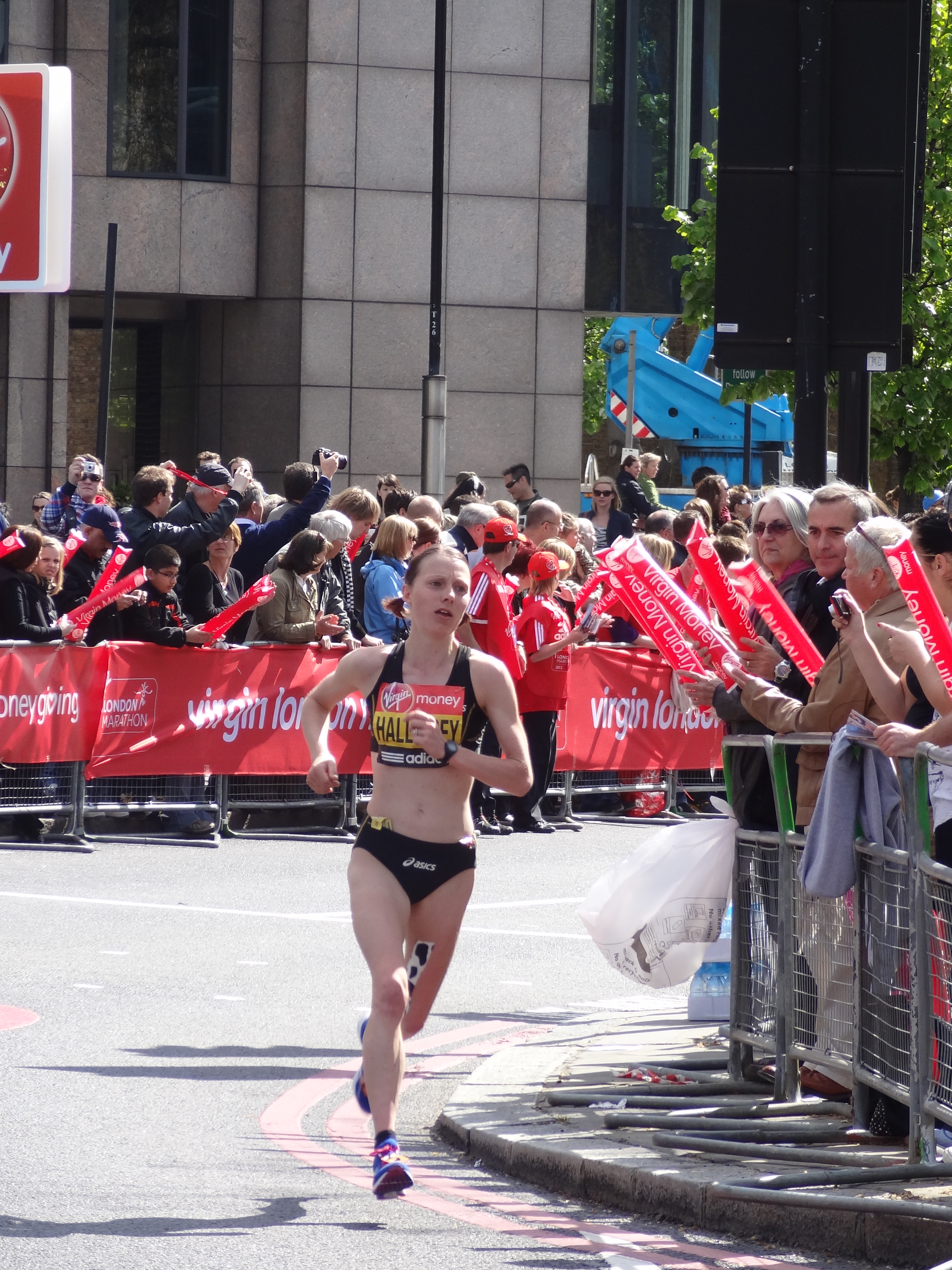
In the women's race, Clare Hallisey (left) finished 11th, however, her time of 2:27.44 was just under the women's Olympic "A" standard of 2:28 & was also faster than the time set by Jo Pavey earlier this year, which had previously placed her in pole position to take Team GB's third Olympic spot in the women's marathon this summer.
One aspect that was dramatically apparent, was the ease at which the leading elite runners seemed to glide over the asphalt, whereas many of the lower-placed elite finishers & a large proportion of the sub-elite runners seemed to be fighting with their biomechanics & wrestling their limbs to get from A to B. It was no surprise that so many were strapped, taped or limping by the time the 22nd mile came into view, but it reinforces the fact that performance requires more than just training, demanding a massive amount of heart & guts to take on & beat the beast of the marathon.
Relating these observations to my earlier blog on tendon pathologies, another critical component of the objective assessment that the clinician must include, is an analysis of the athlete's kinematics.
Azevedo et al (2009) found that in a study
of the biomechanics of 21 runners with Achilles tendinopathy, knee range of
movement between heel strike & midstance was significantly reduced in
comparison to a case matched cohort of 21 injury-free runners. This was associated with reduced tibialis
anterior pre-activation, in addition to reduced rectus femoris & gluteus medius activation
immediately post-heel strike.
Azevedo, L.B. et al (2009). Biomechanical variables associated with Achilles tendinopathy in runners. Br J Sports Med, 43: p288-292
Reduced relative activation of the tibialis
anterior in the 100ms prior to heel strike could indicate an increase in tendon
loading during initial contact, as the pre-stiffening of the tendon-muscle
complex does not occur, thus affecting the ability to absorb the impact forces.
Rectus Femoris acts eccentrically during
early stance phase to restrain the movement of the tibia as the knee flexes
& is an important muscle for shock absorption during this phase of gait. Grau et al (2008) noted eccentric overloading
of the quads muscle group in a cohort of runners with patellar tendinopathy. This could be secondary to an underlying weakness in the quads muscle group, which would result in the premature & prolonged maximal knee flexion
on ground contact. This would in turn lead to decreased speed of hip extension & subsequent
increased speed of knee flexion, which would then be uncontrolled if the knee extensors demonstrated weaker concentric capabilities - a classic viscous circle!
Grau, S. et al (2008). What are causes & treatment strategies for patellar-tendinopathy in female runners? J of Biomechanics, 41(9): p2042-2046
Gluteus medius stabilises the hip with
respect to the thigh in the early stance phase.
Donoghue et al (2008) reported a decreased ability to maintain hip
abduction in early stance phase in subjects with chronic Achilles
tendinopathies compared to a control group.
Grau et al (2008) also reported increased adduction in frontal plane
movement in a cohort of subjects experiencing patellar tendinopathy, which
could be a co-contributor to a change in patellar tendon load already
influenced by a relative premature maximal tibial internal rotation.
Donoghue, O.A. et al (2008). Lower limb kinematics of subjects with chronic Achilles tendon injury during running. Res in Sports Med, 16(1): p23-38
Foot pronation is another kinematic variable that warrants investigation. Grau et al (2008) reported an increased velocity of foot pronation in subjects presenting with patellar tendinopathy, whilst studies investigating Achilles tendinopathy have noted an association with excessive foot pronation (Donoghue et al, 2008; McCrory et al 1999; Ryan et al, 2009).
McCrory, J.L et al (1999). Etiological factors associated with Achilles tendinitis in runners. Med & Sci in Sports & Ex, 31(10): p 1374-1381
Azevedo et al (2009) found a trend of reduced Peroneus Longus activity during the first 100ms after contact in a cohort group suffering from Achilles tendinopathy, which they hypothesised was related to decreased joint stiffness & lateral ankle joint stability, given that similar findings have been reported in groups with functional ankle instability in walking.
Richards et al (2002) conducted a small
study looking at characteristics of ankle joint kinematics that may have had a causative
effect on patellar tendinopathies & suggested that high ankle
inversion/eversion moments, high external tibial rotation & ankle
plantarflexion moments, high ground reaction forces & high rates of
extensor moments around the knee may all have a contributory role to play. However, the experimental cohort was only n =
10, with no matched control group & the study was not a longitudinal
prospective study, therefore it is impossible to determine whether the findings
were the cause or effect of the patellar tendinopathy. As such the study just highlighted kinematic
idiosyncrasies that warrant further investigation.
Richards, D.P. et al (2002). Relation between ankle joint dynamics & patellar tendinopathy in elite volleyball players. Am J Sports Med, 12(5): p266-272
Hopefully, that has either provided some more insight into the components of the objective examination when assessing athletes with tendon pathology, or at least reminded you of why those of you clinicians do what you do! As ever, please feel free to leave comments in the box below if you want to contribute more to the blog. In the meantime, keep on running!